A Systematic Comparison of Alpha-Synuclein Seed Amplification Assays for Increasing Reproducibility
Seed amplification assays (SAAs) enable ultrasensitive detection of misfolded α-synuclein across biofluids and tissues. Yet, heterogeneity in protocols limits cross-study comparability and clinical translation. Here, we review α-synuclein SAA methods and their performance across various biological matrices. We catalog key variables (e.g., human recombinant α-synuclein variants, buffers, salts, beads, shaking, incubation temperature) and outline pre-analytical factors that shape outcomes.
Click here to read the full article →
Relationship between locus coeruleus and slow-wave sleep in aging and Alzheimer's disease

Sleep disruption, particularly loss of slow-wave sleep (SWS), is common in Alzheimer’s disease (AD), but its neurobiological underpinnings remain unclear. We investigated whether locus coeruleus (LC) integrity relates to SWS across the AD continuum and whether sex and perivascular spaces (PVSs) modify these associations.
Click here to read the full article →
Brand-New Biomarker for Alzheimer’s: Circulating, Circular RNA
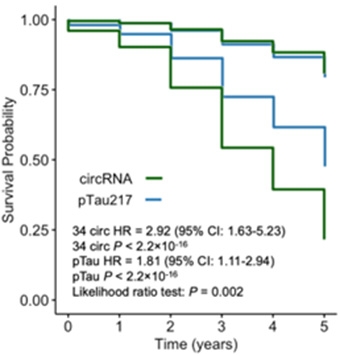
Ring-shaped RNAs floating in a person’s bloodstream might mean bad news for their brain. At the 18th Clinical Trials on Alzheimer’s Disease, held December 1–4 in San Diego, Bridget Phillips, a graduate student in the laboratory of Carlos Cruchaga at Washington University in St. Louis, reported that a select panel of these durable RNA loops can pick out who is at risk of developing Alzheimer’s symptoms even better than does p-tau217. A clinical blood test based on this approach is in development.
Click here to read the full article →
Latest From GNPC: APOE4 Ages Astrocytes, Cripples Energy Metabolism
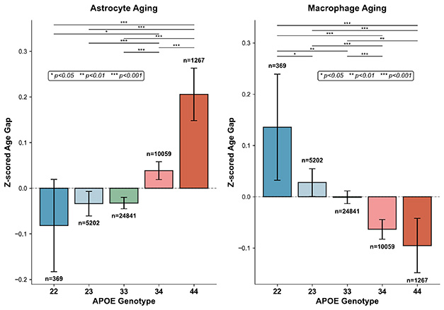
For all the reams of previous research on APOE, scientists are still turning up fresh insights on how its alleles affect biology and disease risk. At the 18th Clinical Trials on Alzheimer’s Disease conference, held December 1-4 in San Diego, three speakers detailed the latest findings gleaned from the Global Neurodegeneration Proteomics Consortium. This large dataset comprises proteomic analyses of 40,000 blood and cerebrospinal fluid samples from more than 20 aging and neurodegenerative disease cohorts.
Click here to read the full article →
ApoE4 Sparks Neuronal Energy Crisis
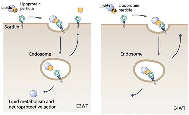
The APOE4 allele heightens the risk of Alzheimer’s disease in several ways. Here’s a new one: It may deprive neurons of a crucial fuel. In the October 16 Nature Metabolism, scientists led by Thomas Willnow at Aarhus University, Denmark, reported that the E4 version of the lipoprotein blocked neurons’ ability to take up polyunsaturated fatty acids via the sorting receptor sortilin. Short on these oils, the cells’ metabolic gears ground to a halt, particularly at synapses. There, mitochondria lost the ability to utilize long-chain fatty acids as fuel.
Click here to read the full article →
Does ‘Runaway Aggregation’ Spell the End for Neurons?
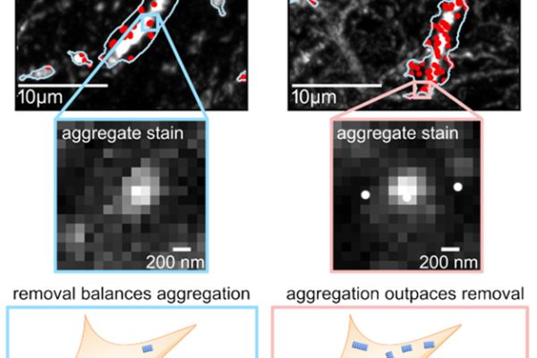
How long can someone bail water before the boat tips over? That depends on two opposing factors: the rush of water, and the fitness of the bailer. A similar push-and-pull may take place in neurodegenerative proteinopathies, where disease hinges on a balance between the production of aggregates, and how efficiently cells can gid rid of them. This is the upshot of a preprint posted October 30 on bioRxiv.
Click here to read the full article →
Does AD Risk Gene INPP5D Leave Lysosomes With a Leak?
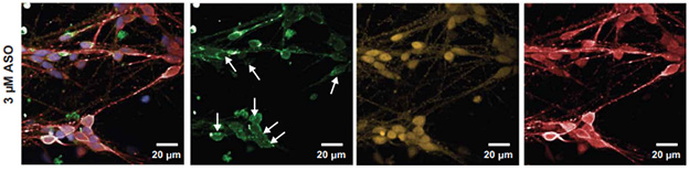
Common variants in the phosphoinositol phosphatase INPP5D are linked to an increased risk of late-onset Alzheimer’s disease, but no one is quite sure why. Recent work suggests that depleting INPP5D in microglia activates the inflammasome and spurs secretion of proinflammatory cytokines. Scientists now blame faulty lysosomes. In a preprint uploaded to bioRxiv on October 27, Tracy Young-Pearse at Brigham and Women’s Hospital, Boston, and colleagues claim that without INPP5D, lysosomes in microglia spring leaks, spilling proteases into the cytosol and setting off inflammatory signaling.
Click here to read the full article →
Waste Products of Gut Bacteria Said to Trigger Brain Aging
The brain gets the spotlight when it comes to how it ages but, behind the scenes, the gut microbiome also transforms over time. As microbial communities shift, so do their metabolites—which can leak into the bloodstream. Is that a problem? Some scientists think yes. In a preprint uploaded to bioRxiv on October 5, Stanford University’s Ami Bhatt and colleagues identified gut-derived metabolites that changed gene expression in the human brain. The findings close a gap in understanding the gut-brain axis by tying metabolites to altered energy metabolism, chromatin structure, and RNA splicing, all of which also change in neurodegenerative diseases.
Click here to read the full article →
Seeing Is Knowing: Introducing the Digital Neuropathology Collection
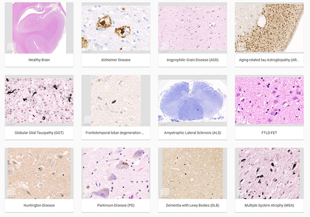
Amyloid-β and tau deposits are visible, in the aggregate, on PET scans, but what kind of havoc do these images reflect at the cellular and molecular level? And what do the deposits even look like, exactly? Neuropathologists know, but to Jochen Herms, Ludwig-Maximilians-University, Munich, this specialty scientist may be a dying breed. “As more and more people work on PET and fluid markers, we may be losing sight of the underlying pathology at the microscopic level,” Herms told Alzforum. With the rise of omics techniques, fewer students and postdocs are trained in neuropathology of disease, much less choose a career in this field.
Click here to read the full article →
New NLRP3 Inhibitor Acts In Brain to Melt Obesity
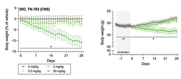
Whether a person eats a cookie or devours the whole box could come down to the mood of microglia in their hypothalamus. Blocking activation of the region’s NLRP3 inflammasome could keep excessive munchies in check, according to preclinical findings posted on bioRxiv October 9.
Click here to read the full article →
Could Taking Down a DNA Repair Protein Stave Off Huntington’s?
At birth, people who carry a string of more than 40 CAG repeats within the first exon of the huntingtin gene are all but destined to develop Huntington’s disease. Yet, recent studies are converging on the idea that the number of inherited repeats is not always sufficient to manifest the disease. Rather, it is their subsequent expansion within subsets of neurons, some of which ultimately harbor hundreds of repeats, that ignites the devastating neurodegenerative disorder. Therein lies an opportunity to stop it, according to scientists led by Sarah Tabrizi of University College London. In the February 12 Science Translational Medicine, they reported that taking down MSH3, a DNA repair protein, nipped CAG expansion in the bud.
Click here to read the full article →
Do Specialized Glycoproteins Prop Up Blood-Brain Barrier?
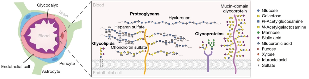
The blood-brain barrier weakens during normal aging and in neurodegenerative disease, but the reasons are not fully understood. In the February 26 Nature, scientists led by Tony Wyss-Coray and Carolyn Bertozzi at Stanford University, California, venture a suggestion. In mice, they found that mucin-domain glycoproteins, components of the carbohydrate meshwork that lines the interior of brain blood vessels, wane with age. In young mice, thinning out these sugary, bottlebrush-shaped proteins made brain blood vessels leak. Conversely, promoting glycosylation of the proteins in old mice firmed up the blood-brain barrier. This even helped prevent memory slippage.
Click here to read the full article →
Big Data Insights: Blood Signatures of Cognitive Decline, Aging, APOE4
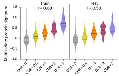
In the “omics” era, where bigger is better, the Global Neurodegeneration Proteomics Consortium is making a splash. To date, this two-year-old initiative led by Gates Ventures and Johnson & Johnson has assembled data from more than 40,000 biofluid samples from 23 neurodegenerative disease cohorts, and it continues to grow. The V1 version of the dataset, comprising 35,056 of those proteomes, was released to researchers July 15 on the Alzheimer’s Disease Data Initiative’s AD Workbench.
Click here to read the full article →
Sniffing Out Early Signs of Parkinson’s Disease
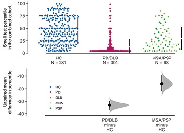
Do quick scratch-and-sniff scents have a bigger purpose than magazine perfume ads? A new study led by Juan Li and Michael Schlossmacher, Ottawa Hospital Research Institute, Toronto, and Brit Mollenhauer, University Medical Center Goettingen, Germany, argues that brief olfactory tests deserve a slot in the PD/DLB diagnostic tool kit. By analyzing three case-control studies, they found seven scents from two popular smell tests—the 16-scent Sniffin’ Sticks Identification Test (SST-ID) and the 40-scent University of Pennsylvania Smell Identification Test (UPSIT)—that distinguished PD/DLB patients from healthy controls as well as did the full-length exams.
Click here to read the full article →
Will Keeping HDLs Lipidated Also Keep Alzheimer’s Away?
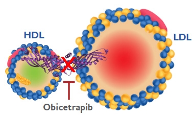
Statins do not help prevent Alzheimer’s disease, but another cholesterol-controlling drug just might. Obicetrapib blocks cholesteryl esters being transferred from the “good” high-density lipoproteins to the “bad” low-density variety. In other words, it boosts HDL-cholesterol at the expense of LDL-cholesterol. In a subset of participants in a large Phase 3 cardiovascular disease trial, the drug also tempered the rise of plasma p-tau217, a marker of amyloid and tau pathology. Obicetrapib even took a bite out of plasma neurofilament light, which flags neurodegeneration but is difficult to shift therapeutically, said Philip Scheltens, EQT Life Sciences, Amsterdam. Scheltens presented the findings at the Alzheimer’s Association International Conference, held July 27-31 in Toronto.
Click here to read the full article →
Coming Soon: Africa’s First Large-Scale Alzheimer’s Datasets
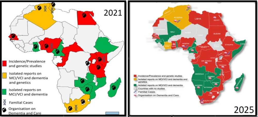
Rufus Akinyemi marked a milestone at AAIC 2025, held 27-31 July in Toronto. “This is the very first time the conference has dedicated multiple sessions to dementia research in Africa,” he said. Indeed, there is more dementia research in Africa happening year-on-year. Akinyemi and Adesola Ogunniyi, both at the University of Ibadan, Nigeria, presented an update to their 2021 review paper summarizing the state of knowledge about dementia in Africa, which had shown but a sparse patchwork of studies across the continent (Akinyemi et al., 2021). Now, their revised map features many more countries contributing to the evidence base, with only a handful of African nations still lacking a dementia publication (image below). “We are very happy with the progress being made,” Akinyemi said, to applause.
Click here to read the full article →
TfR, No Longer Lone Star? New Shuttles Use Other Keys to Unlock Brain
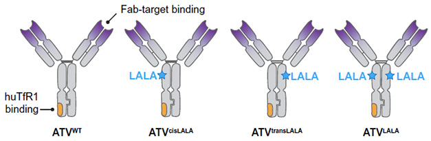
The highly selective border lining the brain’s vasculature is the Achilles’ heel of any large therapeutic molecule aiming for targets in the brain, anti-Aβ antibodies included. Not only do fewer than 1 percent of these molecules manage to cross from the blood into the brain, but those that do tend to get snagged by clumps of vascular amyloid just on the other side, where they can instigate inflammation and dangerous brain swelling.
Click here to read the full article →
Fluid Marker for Parkinson’s Prognosis: Are We There Yet?
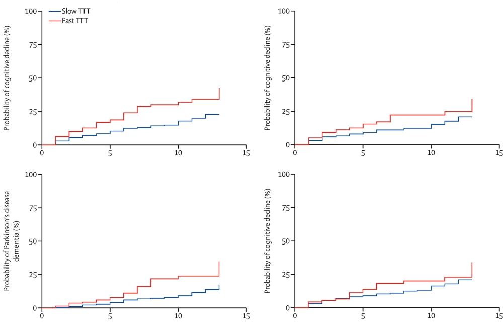
Parkinsonian disorders are difficult to diagnose. Clinical symptoms often overlap with criteria for other diseases, and clinicians can only make a definitive diagnosis postmortem. Now, a study published in the July 2025 issue of Lancet Neurology suggests scientists may be closer to a cerebrospinal fluid test that can distinguish people with different forms of parkinsonism. Researchers led by Edwin Jabbari, at University College London, report that α-synuclein seed amplification assays help differentiate Parkinson’s disease from Progressive Supranuclear Palsy and predict whether patients will develop cognitive decline.
Click here to read the full article →
New Type of Aβ42 Fibril Discovered in 83-Year-Old Man
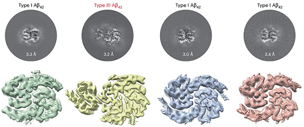
Thanks to cryo-electron microscopy, scientists have solved the structure of amyloid and tau fibrils—the hallmark protein pathologies of Alzheimer’s disease, and at the atomic level. As for Aβ42, they have described two types of fibrils. Type I, found in sporadic AD, comprise two identical S-shaped protofilaments that stack and twist together. Type II, found in familial AD, have a similar S-twisted protofilament that stacks in a different configuration (Jan 2022 news). Now, Wenying Qiu of Peking Union Medical College, Beijing; Cong Liu of Fudan University, Shanghai; and Dan Li of Shanghai Jiao Tong University report a third amyloid fibril, Type III, in the June 10 Nature Chemical Biology. Found postmortem in the soluble fraction of a brain from an 83-year-old man, it comprised three protofilaments twisted together.
Click here to read the full article →
A Tale of Two T Cells—Slowing Tau Spread, Shredding White Matter
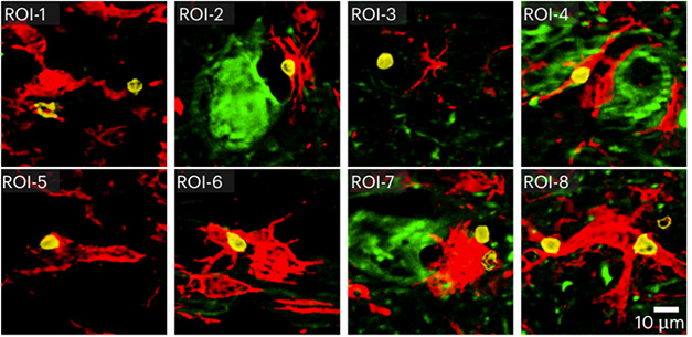
Meet the Jekyll and Hyde of the immune system: activated CD8⁺ T cells. On June 24 in Nature Immunology, researchers led by Dorian McGavern at the National Institutes of Health introduce a granzyme K–wielding subset that rush to distressed microglia in a mouse model of tauopathy and slow down the spread of phosphorylated tau and neurological decline. Just a month earlier, however, Mikael Simons’ lab at the Technical University of Munich had revealed a sinister side of T cells. On May 22 in Nature Neuroscience, they showed that in older, wild-type mice, inflammatory microglia summon a cadre of cytotoxic T cells that ravage myelinated axons in the optic nerve. So, are they friend, foe—or both?
Click here to read the full article →
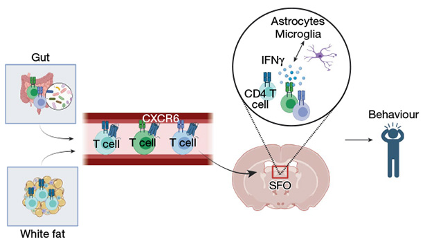
T Cells from the Gut Occupy a Niche in Human and Mouse Brain
Click here to read the full article →
Alpha-synuclein misfolding as fluid biomarker for Parkinson’s disease measured with the iRS platform
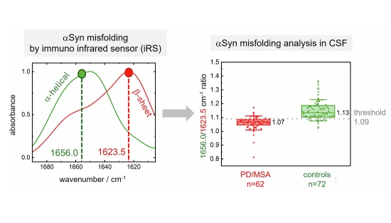
A new fluid biomarker for Parkinson’s disease (PD) was developed, offering a high-accuracy biological classification, potentially at early stages.
A discovery and a validation cohort consisting of 134 individuals was analyzed, using α-synuclein misfolding in CSF as a biomarker, measured by the iRS (immuno-infrared sensor.
The iRS shows 97% sensitivity and 92% specificity in classifying PD/ multiple system atrophy (MSA) versus controls.
The structure-based biomarker enables continuous monitoring of the disease progression.
Click here to read the full article →
Can a Pen Powered by AI Detect Parkinson’s?
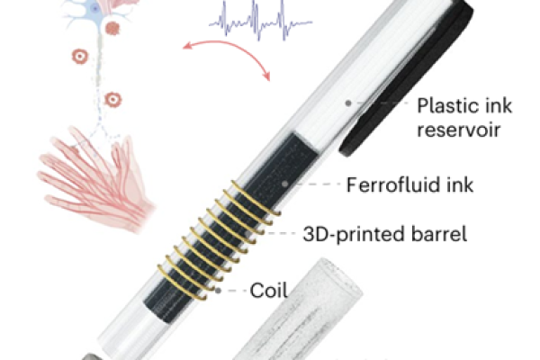
Parkinson’s disease (PD) is the second most common neurodegenerative disorder, after Alzheimer’s. Clinicians often use the mnemonic TRAP to recall its four key motor symptoms: tremor, rigidity, akinesia/bradykinesia, and postural instability. Beyond these hallmarks, additional symptoms, including micrographia—a characteristic reduction in the size of a person’s handwriting, can provide diagnostic clues. Now, scientists led by Jun Chen at the University of California, Los Angeles, have developed a way to enhance PD diagnosis through handwriting analysis. As reported in Nature Chemical Engineering, June 2, they developed a pen that captures writing motions analyzed with artificial intelligence algorithms. In a small pilot trial, the pen diagnosed PD with 96 percent accuracy.
Click here to read the full article →
Could Monocyte-Derived Microglia Cause Some Cases of Parkinsonism?
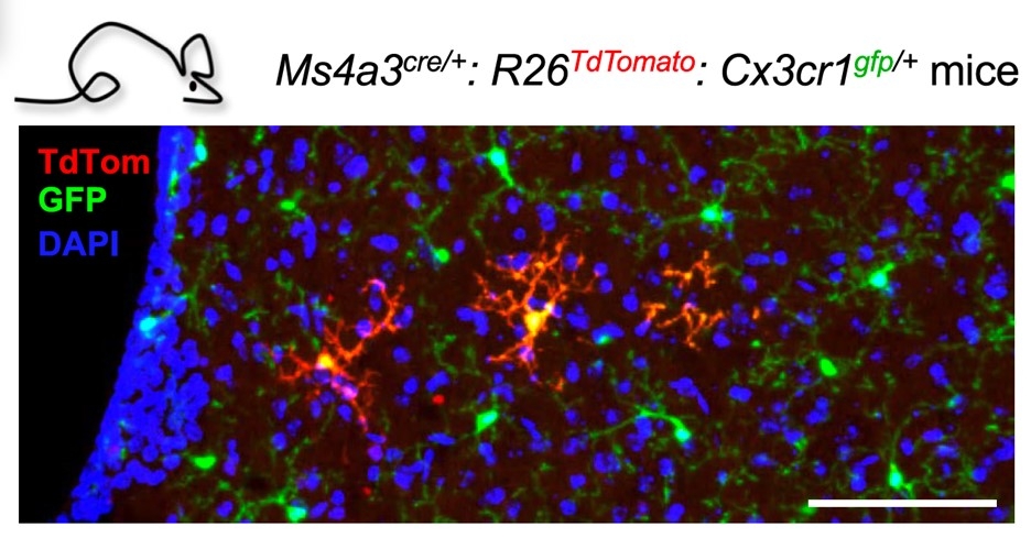
Microglia in the brain’s parenchyma arise from macrophage progenitors in the embryonic yolk sac (Ginhoux et al., 2010), and are thought to self-renew locally during a mammal’s lifespan, without macrophages from the hematopoietic system adding to their numbers.
Click here to read the full article →
Do PERK’d Up Astrocytes Slow Amyloid Clearance?
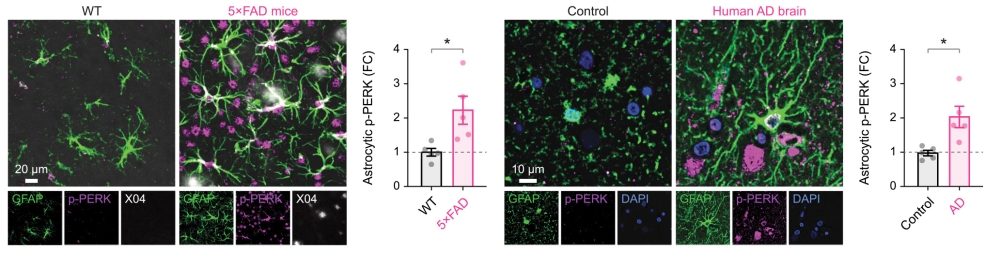
Cells feature an array of biological cascades to respond to stressors such as protein aggregates, viruses, and DNA damage. When these pathways are activated too much or for too long, things can go awry. This may be the case for the unfolded protein response (UPR) in people with Alzheimer’s disease. In Neuron, May 21, scientists led by Guang Yang at Columbia University in New York and Zhongcong Xie at Massachusetts General Hospital reported that, in postmortem human brain and in mouse models of amyloidosis and tauopathy, the UPR—specifically the branch driven by protein kinase RNA-like ER kinase (PERK)—ramps up explicitly in astrocytes. This worsened pathology, in part by impeding glymphatic clearance, a waste-management system in the brain. Knocking out PERK in astrocytes, or blocking it, restored clearance, reduced amyloid pathology, and improved learning and memory, the authors report.
Click here to read the full article →
Astrocytes, the Gatekeepers of Norepinephrine Signaling in the Brain
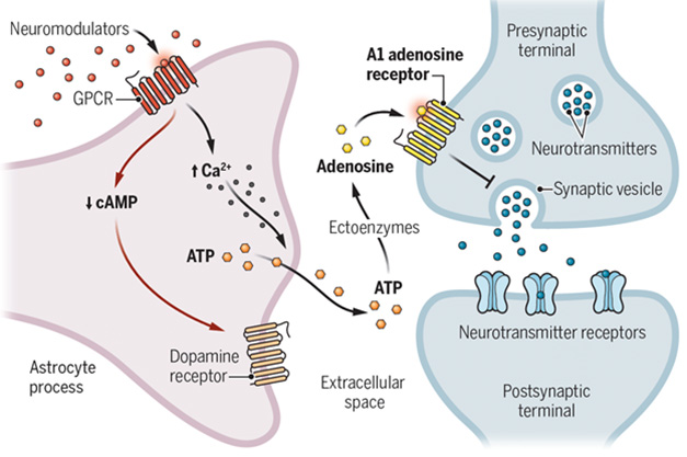
As a neuromodulator, norepinephrine is a powerful force in the brain. We all know it orchestrates rapid changes in behavior such as the fight-or-flight response. Fewer people know that astrocytes appear to be the ones calling norepinephrine’s shots. This is the gist of three papers published May 15 in Science. Using zebrafish, fly, and rodent models, the studies converge on the idea that engagement of adrenergic receptors on astrocytes profoundly influences the fine-tuning of thousands of synapses within their grasp. In contrast, at least for some responses, adrenergic receptors on neurons were dispensable.
Click here to read the full article →